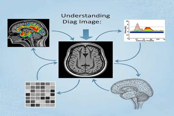
When you hear the term “diag image” (short for diagnostic image), it may sound technical, but it’s really just a fancy way of saying medical picture. These are the images doctors use—like X-rays, CT scans, MRIs, and ultrasounds—to peek inside the body and figure out what’s going on. From spotting a broken bone to tracking the progress of a treatment, diagnostic images are at the very heart of modern medicine.
In this guide, I’ll walk you through what diag images are, how they’re made, why they matter, and how healthcare professionals actually use them. Think of this as your crash course in medical imaging—explained simply, step by step.
What Exactly Is a Diag Image?
At its core, a diag image is just a visual representation of the inside of your body. These images are produced by specialized machines and then interpreted by trained doctors (often radiologists).
Why Are They So Important?
Diag images help with:
-
Diagnosing health conditions – like fractures, tumors, or infections.
-
Monitoring treatment progress – checking if a therapy or surgery is working.
-
Guiding medical procedures – making surgeries or interventions safer and more accurate.
Without these images, healthcare would be a lot more guesswork and a lot less science.
Common Types of Diagnostic Images
Different imaging techniques show different things. Here are the most common types you’ll hear about:
-
X-ray – Best for bones and dense tissues. Think broken bones or chest exams.
-
CT (Computed Tomography) scan – Combines multiple X-rays to create cross-sectional views. Perfect for detailed looks at organs and complex injuries.
-
MRI (Magnetic Resonance Imaging) – Uses magnets and radio waves to capture soft tissue details, like muscles, brain, and spinal cord.
-
Ultrasound – Uses sound waves for real-time imaging. Commonly used in pregnancy, but also for checking organs and blood flow.
-
PET (Positron Emission Tomography) – Shows metabolic activity in tissues, often used in cancer diagnosis.
Each method has its strengths, and doctors choose the right one depending on the situation.
How Are Diag Images Created? (Step-by-Step)
Here’s a simplified look at how medical imaging works:
-
Preparation – Patients may need to fast, remove jewelry, or wear a hospital gown.
-
Positioning – You’ll be placed on an imaging bed or table in the correct position.
-
Image Capture – Depending on the machine, it could involve X-ray beams, magnetic fields, sound waves, or tracers.
-
Data Recording – Special detectors pick up signals from the body and send them to a computer.
-
Reconstruction – The computer processes the raw data into a clear, readable image.
-
Interpretation – A radiologist reviews the image, compares it to previous scans, and writes a report for your doctor.
Pretty amazing, right? In just minutes, these machines can reveal what’s happening inside your body without a single incision.
File Formats: How Diag Images Are Stored
Not all medical images are saved the same way. Here are the main formats:
-
DICOM (Digital Imaging and Communications in Medicine) – The healthcare standard. Stores both the image and essential patient info like name, age, and scan details.
-
JPEG/PNG – Great for sharing or publishing, but not ideal for diagnosis since they may lose quality and metadata.
-
TIFF – High-quality, lossless format, though not as widely used in medical systems.
Key takeaway: DICOM is the gold standard in healthcare because it keeps both image quality and critical patient data intact.
Best Practices for Handling Diag Images
Healthcare professionals take a lot of precautions when working with diagnostic images. Some best practices include:
-
Protect patient privacy – Secure storage and anonymization when sharing.
-
Use PACS (Picture Archiving and Communication System) – A centralized system for storing and retrieving images.
-
Consistent labeling – Including patient ID, date, and scan type.
-
Easy retrieval – Organizing by modality (CT, MRI, ultrasound) and body part.
These steps ensure doctors can access accurate, secure images whenever they need them.
How Doctors View and Interpret Diag Images
If you’ve ever wondered what happens once your scan is taken, here’s a breakdown:
-
Opening the file – Using specialized DICOM viewers like OsiriX, RadiAnt, or Horos.
-
Adjusting settings – Changing brightness, contrast, or zoom to highlight details.
-
Identifying normal anatomy – Recognizing healthy bones, tissues, and organs.
-
Spotting abnormalities – Looking for signs of fractures, tumors, fluid buildup, or blockages.
-
Comparing past images – Tracking changes over time (e.g., healing fractures or tumor shrinkage).
-
Reporting findings – Writing a detailed report with impressions and recommendations.
It’s a blend of technology and human expertise.
Also Read : What Are the Different Types of Massage?
Real-Life Example
Imagine you sprain your wrist badly. An X-ray can confirm whether it’s just a sprain or an actual fracture. If the X-ray doesn’t show much but pain persists, your doctor may order an MRI to check for ligament damage. This layered approach is how diag images save patients from unnecessary treatments and help doctors make precise decisions.
FAQs About Diag Images
Q1: What does “diag image” mean?
It’s short for diagnostic image, which refers to medical pictures like X-rays, MRIs, and CT scans.
Q2: Which scan is best for bones?
X-rays are the go-to for bones, while MRIs and CT scans are better for soft tissues and organs.
Q3: Why is DICOM the most used format?
Because it includes both the image and key patient details, ensuring accuracy and compatibility across healthcare systems.
Q4: Can JPEG replace DICOM?
Not really. JPEGs are fine for presentations but lose metadata and detail, making them unsuitable for official diagnoses.
Q5: How can I view a DICOM file at home?
You can use free viewers like Horos (Mac) or Weasis (cross-platform) to open them.
Q6: Are diag images safe?
Yes—though some, like X-rays and CT scans, involve radiation, doctors only recommend them when benefits outweigh risks.
Why This Matters to You
Diagnostic imaging isn’t just for hospitals and clinics—it touches nearly everyone’s life at some point. Whether it’s checking for a fracture, monitoring a chronic condition, or ensuring your baby is healthy during pregnancy, diag images are the silent heroes of healthcare.
Conclusion
Diag images may sound technical, but at the end of the day, they’re simply medical pictures that help doctors see inside your body safely and accurately. From X-rays to MRIs, these tools guide everything from simple diagnoses to life-saving treatments.
By understanding how diag images work, how they’re stored, and how professionals interpret them, you get a better appreciation of just how critical they are in keeping us healthy.
Next time your doctor recommends a scan, you’ll know exactly what’s going on behind the scenes—and why it matters.

